

Leslie B. Arey Professor, Department of Cell and Developmental Biology, Northwestern University Feinberg School of Medicine, Chicago
Could you please tell us about your research field?
We study the dynamics and functions of the cytoskeleton in eukaryotic cells. Our focus is particularly on the roles of microtubules and intermediate filaments. To investigate these structures, we use different models, including cells and tissues from Drosophila melanogaster and cultured mammalian cells. Through our research, we aim to uncover how cytoskeleton components contribute to cellular processes, for instance neurite outgrownt and polarity determination, and overall cell function, such as motility, cell signalling, transport of organelles and other components. More details about our work can be found on our lab’s website here.
What biological questions within your project can be addressed using fluorescence microscopy?
All of our experiments involve imaging the cytoskeleton and other cellular organelles in live cells, utilizing a range of fluorescent tags to visualize these components. By applying different types of fluorescent markers, we can closely monitor and track the behavior of cytoskeletal structures and their interactions with other cellular elements. This approach allows us to observe how the movement and dynamics of these cytoskeletal components contribute to changes in cell morphology and influence cellular motility. It is precisely by analyzing these live-cell interactions and movements, we gain insights into the underlying mechanisms that govern cellular functions and responses.
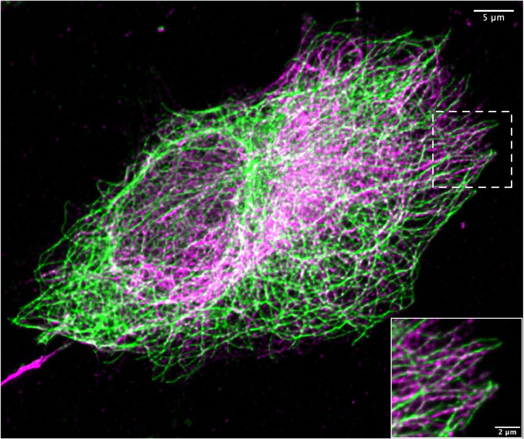
Endogenous microtubule (green) and vimentin (magenta) organization in RPE cells visualized by immunofluorescence staining. The image was acquired using the CrestOptics DeepSIM-X-light (#S8002) mounted on the Nikon Eclipse Ti microscope stand, equipped with a Plan Apo λ 60×/1.4 N.A. oil immersion objective lens.
cellular functions and responses”
When you had to buy new microscopy equipment, how did you choose the CrestOptics DeepSIM system?
The CrestOptics DeepSIM system perfectly complements the capabilities of the existing microscopes in our lab. DeepSIM design is optimized for speed and ease of operation, allowing us to efficiently achieve high-quality imaging with minimal effort. The system provides the super-resolution necessary for our research needs, ensuring that we can capture detailed and precise images. Additionally, the DeepSIM system integrates seamlessly with Nikon Elements software, as well as with Nikon microscope stands and optics, creating a cohesive and user-friendly imaging solution that enhances our laboratory’s overall functionality.
Can you please describe your microscope setup equipped with the DeepSIM?
Our microscope setup features the CrestOptics DeepSIM system installed on the Nikon Eclipse Ti stand. This advanced configuration is complemented by a Prime sCMOS camera (Teledyne Photometrics), which ensures high-quality imaging. The entire system is driven by Nikon Elements software, providing a seamless and integrated user experience. This setup enables us to fully utilize the DeepSIM’s advanced imaging capabilities while benefiting from its versatility in combining with different branded devices.
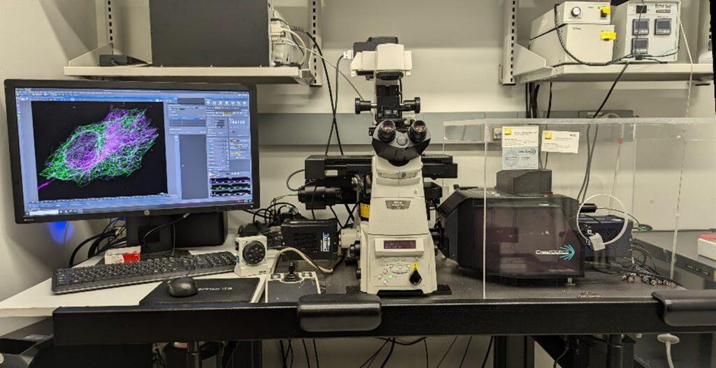
Microscope set-up are located in the Prof. Valdimir Gelfand lab.
“This setup enables us to fully utilize the DeepSIM’s advanced imaging capabilities while benefiting from its versatility in combining with different branded devices.”
How do you see the future of fluorescence microscopy to keep up with innovative biological applications and scientific needs?
The future of fluorescence microscopy is crucial for advancing innovative biological applications and meeting scientific needs. Biological research, particularly in cell biology, is significantly influenced by the available technologies. As such, the development of new microscopy techniques that enhance resolution, speed, and sensitivity of detection is vital. These advancements will enable researchers to observe cellular processes with unprecedented detail and accuracy. Additionally, it is important that these new technologies are user-friendly and accessible to biologists. This convenience will allow researchers to seamlessly integrate advanced microscopy into their workflows, thereby accelerating scientific discoveries. Improved microscopy techniques will be instrumental in deepening our understanding of normal biological processes and how they are disrupted in pathological conditions.

Dr. Bhuvanasundar Renganathan and Dr. Vladimir Gelfand, Department of Cell and Molecular Biology, Northwestern University, Chicago.
We would like to thank Nikon Instruments for providing us with this opportunity and for their continuous support.


