Interview with Dr. Pablo Hernández Varas, Head of Bioimaging Core Leuven, VIB (Flemish Institute for Biotechnology – Vlaams Instituut voor Biotechnologie), Belgium
“I have been working with microscopes for more than 15 years, and I am still passionate about making new biological discoveries with them. As an active scientist I did focus on investigating in detail how cells attach to the substrate, a complex process involved in many physiological functions such as the immune response, but also in pathologies, such as cancer metastasis. I used live cell imaging as well as super-resolution techniques for this”, said Dr. Pablo Hernández Varas, Head of the Bioimaging Core Leuven. After the postdoc, he switched gears, and he has been working in Core Facility environments for the past 8 years. He finds this is a wonderful environment where you can have different microscopy techniques at hand, and you can contribute to many, diverse research projects helping the researchers acquire and understand their imaging data. “I have a more comprehensive view on Bioimaging now than I used to, spanning from spinning disk confocal to single molecule imaging, and I am thrilled every day helping our users to find the right microscopy tool to get the scientific answer they need to push their research forward”, he added.
SPINNING DISK CONFOCAL: fast, gentle, sensitive
We have a very broad user base, as we service two large research centers, the Center for Brain and Disease (CBD) and the Center for Cancer Biology (CCB). That means that we get a bit of everything. Many researchers need standard tools for histopathology, for example, where massive, systematic scanning of hundreds of tissue slides is required. Imagine scanning brain sections looking for signs of Alzheimer’s disease, or a pancreas invaded by a nasty tumour from a mouse model. However, the most used technique is by far confocal, due to its optical sectioning capabilities. These days having three-dimensional context is paramount to understand the biological interactions, and hence being able to acquire sharp Z-stacks and reconstruct the volume of the sample is required. Although we still rely on point scanner confocals as the default go-to tool for volumetric imaging, more and more samples are requiring spinning disk confocal imaging. These come in two main flavours: first, spinning disk is gentler and allows for live cell imaging over time, which is really interesting in order to explore 3D cellular behaviour and dynamic inter-cellular interactions; secondly, imaging large, cleared specimens like brain organoids or tumoral masses requires fast and sensitive imaging systems, and the spinning disk is, in combination with new silicon lenses, unbeatable due to the balance between optical sectioning, imaging depth, speed and ease of use.
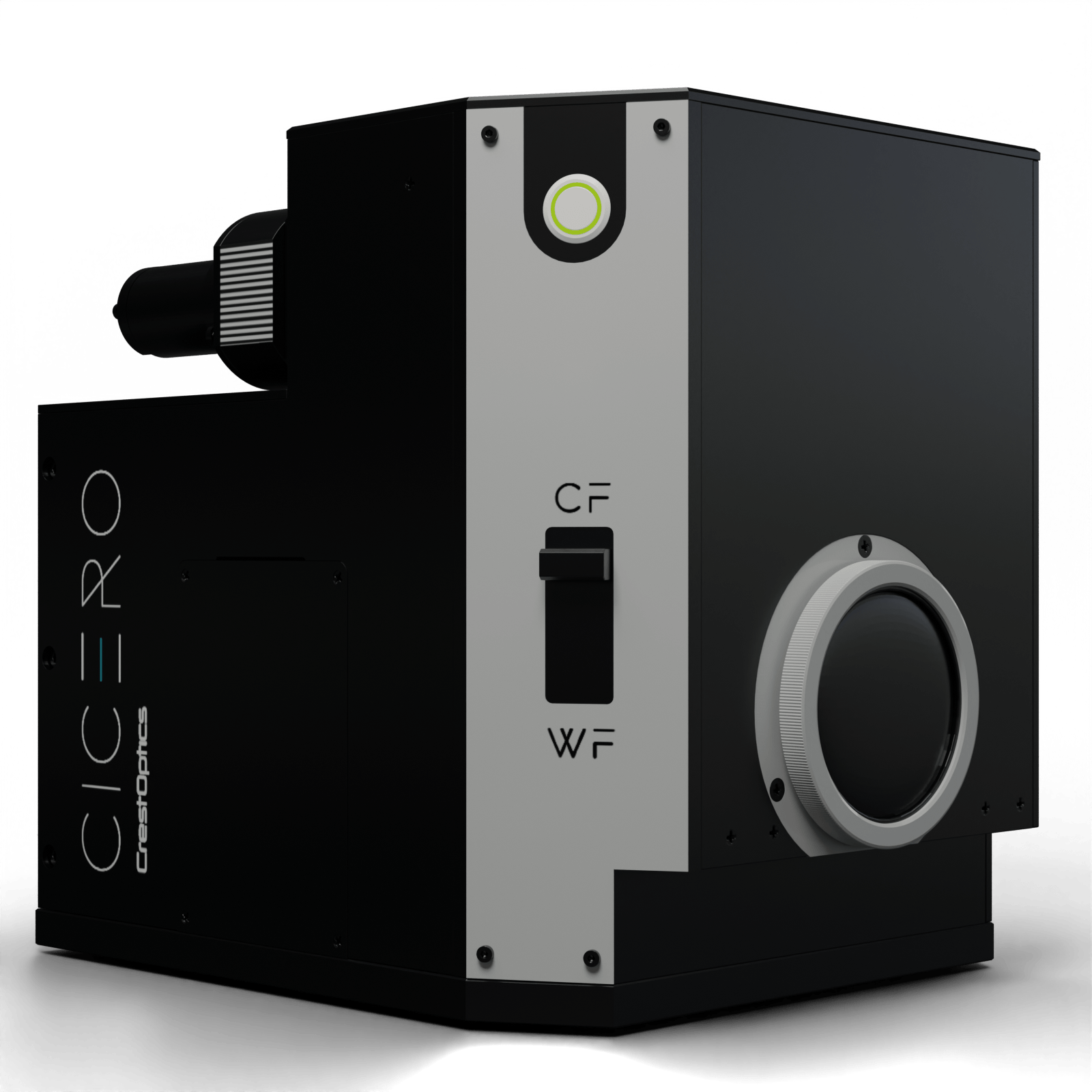
CICERO in one word: easy!
If I must say something about the CICERO spinning disk in one word, that word would be “Easy”! Our experience with the system has been overwhelmingly positive, having excellent support during a simple installation and setup procedure, and having experienced plain sailing after that. We have put the system through a tough test, imaging multiple cleared, large samples, almost continuously, and it has behaved very well, being reliable and robust. Our users were impressed and could use it almost independently after a few minutes. The system has one camera but still it was blazingly fast thanks to its fast-spinning disc and powerful light source, combined with the latest generation sCMOS camera.
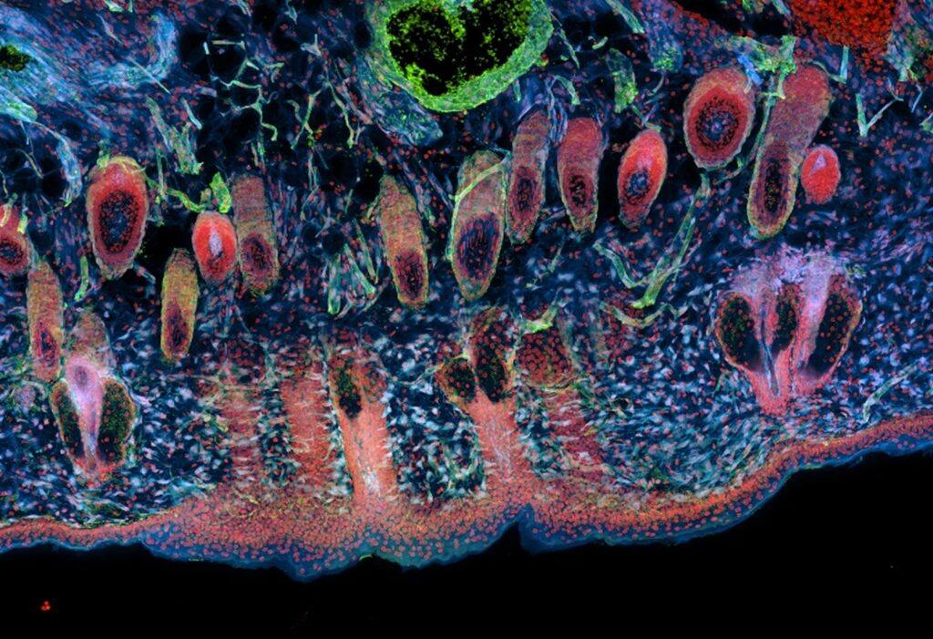
Figure 1: Mouse melanoma allograft model. A Maximum Intensity Projection of a 30 µm thick section acquired with CICERO Spinning Disk is shown. The high-quality sections allow the visualization of the innervation in both tumor and skin tissues.Sample provided by Cecilia Pazzi from the laboratory of Chris Marine (CCB – VIB), imaging by Nikky Corthout (BIC Leuven).
CICERO: enhancing imaging capabilities across various applications
For our researchers, the CICERO will bring speed and gentleness to our confocal acquisitions. So far, point scanning confocals have been the most used tool at the facility. However, scanning one pixel at the time is inherently slow. The beauty of a system such as the CICERO is that the acquisition speed is improved by an order of magnitude, without sacrificing much of the confocality if you choose your lenses correctly. Moreover, and not to be underestimated, the easy of use is remarkable. Just a few clicks and you get a beautiful, optically sectioned image. These two features will be particularly important for users requiring large sample volumetric imaging. Many of the researchers at the Centers we service do clearing of large samples. This could well be a full mouse brain, looking at neuronal connectivity or Alzheimer’s β-amyloid plaque formation, or an invading tumor from a mouse model or even a human patient. For these applications the spinning disk makes a difference. Also, some of our researchers will be looking at dynamic processes, such as organoid growth or tumour formation, which we did thoroughly test. For our researchers, the combination of gentleness (another advantage of spinning disk over conventional point scanning confocals) and speed is a deal breaker, enabling live cell imaging otherwise not possible.
THE EXPERT’S VOICE ON CRESTOPTICS TECHNOLOGY
“We have put the system through a tough test, imaging multiple cleared, large samples, almost continuously, and it has behaved very well, being reliable and robust.”
Dr. Pablo Hernández Varas, Head of Bioimaging Core Leuven, VIB (Belgium)