Unlocking the secrets of virus-host interactions: a look into the SpacVir Team's Research using the CICERO Spinning Disk Confocal
Team SpacVir - Laboratoire de Microbiologie Fondamentale et Pathogénicité (MFP)
CNRS / Université de Bordeaux UMR 5234, France
The spatial and temporal control of virus-host interactions team (SpacVir) is interested in how viruses interfere with cellular processes during virus entry and the priming of viral genomes for gene expression and replication. Our focus is on the spatial and temporal organization of host-pathogen interactions during these processes to understand their principles and identifying potential novel drug targets. We have specialized in the in vivo imaging of viral nucleic acids and viral interactions with host intrinsic defense mechanisms. Our team is headed by a basic scientist and a medical virologist. This unique combination allows us to study fundamental aspects of virus biology and make the link to practical clinical needs. We are also strongly involved in virology teaching at Bordeaux University covering the vast majority of virology-related lectures and practical courses in the medical faculty and the natural sciences faculty.
CICERO: the ease of installation
The SpacVir team required a cost-effective and easy-to-use confocal system to be integrated into an existing imaging setting in our BSL2 laboratory without compromising on image quality. For years, we wanted to be able to track infections in cells with confocal resolution over prolonged periods with minimal phototoxicity. For this purpose, Spinning Disk technology is the best choice. “We chose the CICERO Spinning Disk system from CrestOptics with the help of Marie-George Côme from ImagXcell after she arranged a demo of our existing system“, Dr. Harald Wodrich, Director of Research at MFP-CNRS said. We have a Leica DMi-6000 inverted microscope equipped with an Aura III source from Lumencor and a Hamamatsu Orca Flash 4 Camera embedded into an environmental chamber. We control all our systems through MetaMorph software. The ease of installation, the performance, and the quality of the images acquired immediately appealed to us. With the acquisition of CICERO, we were able to upgrade our acquisition system to a spinning disk confocal at a reasonable cost. We can now carry out confocal acquisitions, especially on living cells infected by replicating viruses over time in complete safety.
THE EXPERT’S VOICE ON CRESTOPTICS TECHNOLOGY
“The ease of installation, the performance, and the quality of the images acquired immediately appealed to us. With the acquisition of CICERO, we were able to upgrade our acquisition system to a spinning disk confocal at a reasonable cost.”
Dr. Harald Wodrich, Director of Research at MFP-CNRS Université de Bordeaux (France)
CICERO: key features assisting researchers
The acquisition and installation of the CICERO Spinning Disk from CrestOptics allowed us to follow infections in reconstituted bronchial epithelia over several days. We also were able to track individual viral genomes at the onset of infections and follow their dynamic distribution. We also used confocal images on fixed samples to identify differences between wild-type and mutated viruses differentiating between their behaviour in single cells vs. complex tissues. We particularly favour time-lapses at multiple wavelengths in confocal mode on infected cells to determine the infection dynamics of viral features at the same time as cellular features. With the CICERO setting, we have started to look again into many cell lines and many mutant viruses that we created throughout the years monitoring viral and host proteins of interest during infections using fluorescent fusion proteins such as GFP, mCherry and tdi-RFP (far-red). The simultaneous use of multiple colours at spinning disk resolution with the benefit of low photo bleaching is great.
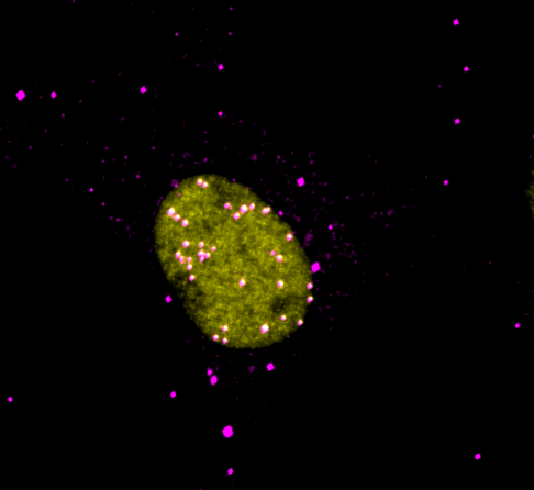
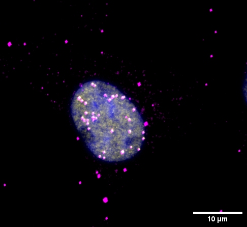
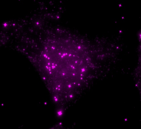
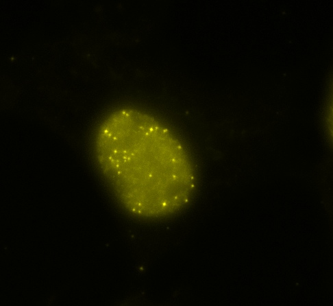
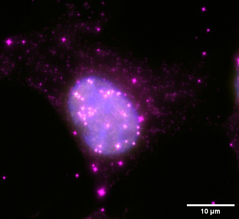
A. WIDEFIELD
CICERO SPINNING DISK CONFOCAL ACQUISITION
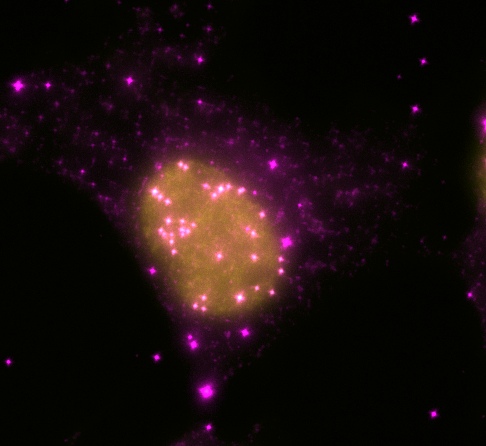
A. WIDEFIELD
CICERO WIDEFIELD ACQUISITION
Figure 1: Comparison between CICERO Spinning Disk Confocal (TOP) and Widefield (BOTTOM) imaging. Evaluation of the nuclear import of Adenovirus 5 genomes upon infection in U2OS cells. Staining: Dots of viral protein VII associated with viral genomes (Total protein VII) is shown in magenta; dots of TAF1ß cellular nuclear factor which associates to viral protein VII upon nuclear import of viral genomes. (Only nuclear protein VII) are shown in yellow; DAPI for nuclei staining is shown in blue.
CrestOptics & ImagXcell: successful stories across France
The productive collaboration between ImagXcell and CrestOptics has generated several successful stories across France. This partnership benefits from the manufacturer’s commitment to innovation, never losing sight of the customer’s application needs and a distributor with over 15 years of expertise in integrating spinning disk solutions. This synergy allows us to provide a cost-effective solution without compromising on performance or image quality. It serves as a prime example of seamless installation and integration of the CICERO Spinning Disk Confocal system into a BSL2 laboratory imaging setup, particularly suitable for laboratories with limited budgets.
In virology, the CICERO Spinning Disk Confocal system has proven to be indispensable. Its capability to capture high-resolution images with minimal phototoxicity has enabled researchers at SpacVir to explore various cell lines and mutant viruses and to track viral infections in real-time, even over extended periods. This has been crucial for research to understand the dynamics of virus-host interactions, notably during viral entry and genome expression. Moreover, the system’s user-friendliness and compatibility with various fluorescent fusion proteins have facilitated simultaneous study of viral and host protein dynamics, offering valuable insights into infection mechanisms. Overall, the CICERO system stands out as the instrument of choice for enhancing virology research capabilities, enabling significant advancements in understanding viral pathogenesis, and exploring potential therapeutic interventions.
Marie-George Côme
Business Development Manager & Founder
ImagXcell, France
CrestOptics extends its gratitude to Nicolas Landrein, Engineer at The MFP – CNRS – University of Bordeaux, for his assistance in the installation of CICERO and support in the interview content. We would also like to express our gratitude to Marie-George Côme (ImagXcell) for giving us this chance and for her contribution.
Subscribe to MicroLife to receive a monthly update on CrestOptics news